
Case Studies
The case studies and images below have been supplied by healthcare experts throughout the country that have seen the value of the enhanced images provided by the use endorectal coils.
» View articles on MRI Prostate Imaging with Endorectal Coils
Interested in viewing our 3.0T Case Studies?
1.5T Case Study 1
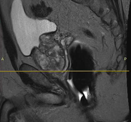
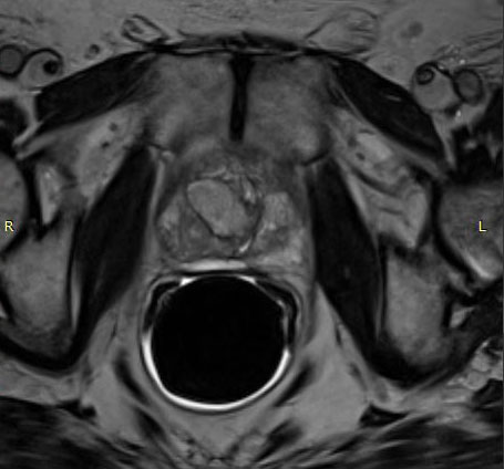
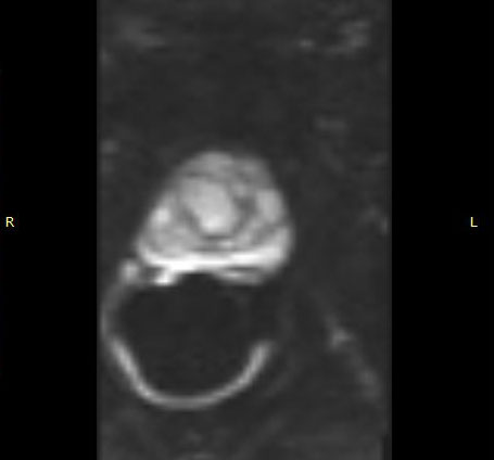
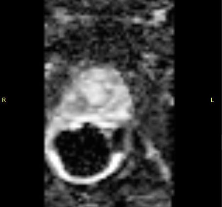
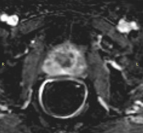
Case courtesy of Crozer-Chester Medical Center
1.5T Case Study 2
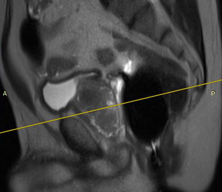
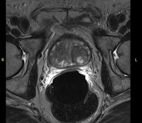
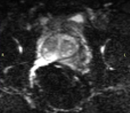
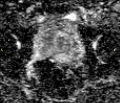
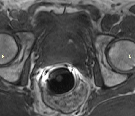
Case courtesy of VA Hospital
1.5T Case Study 3
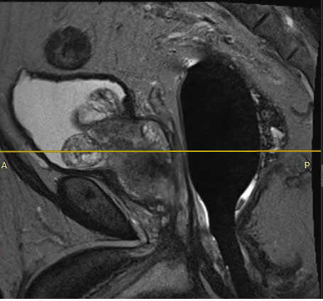
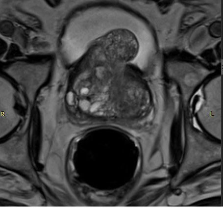
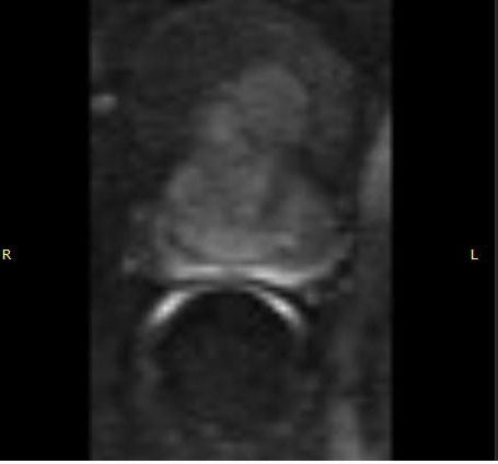
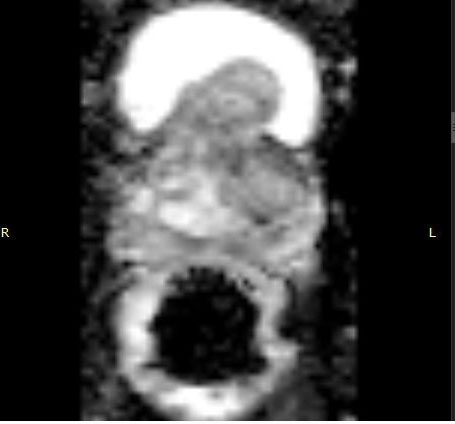
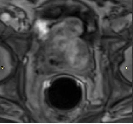
Case courtesy of Crozer-Chester Medical Center
3.0T Case Studies
The case studies and images below have been supplied by healthcare experts throughout the country that have seen the value of the enhanced images provided by the use endorectal coils.
» View articles on MRI Prostate Imaging with Endorectal Coils
Interested in viewing our 1.5T Case Studies?
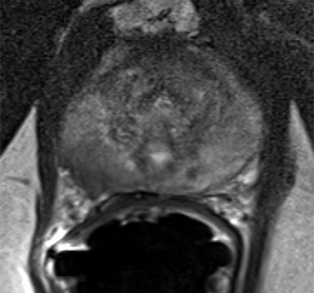
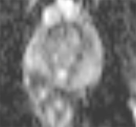
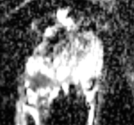
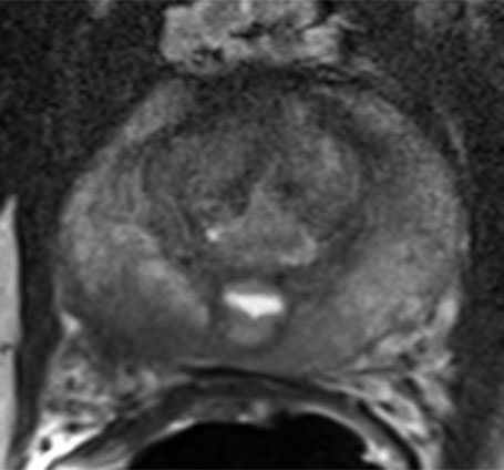
3.0T Case Study 1 – 53 year old male, elevated PSA of 10 ng/mL
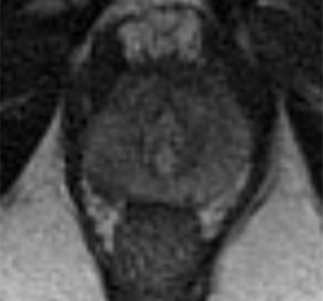
T2w w/o ERC
T2w with ERC
ADC w/o ERC
ADC with ERC
T2W
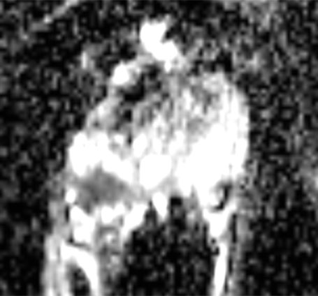
ADC
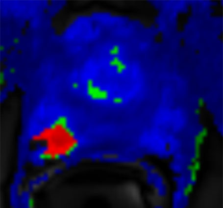
DCE
Case courtesy of Rajan T. Gupta, MD; Duke University Medical Center
3.0t Case Study 2 – 45 year old male, elevated PSA of 8.6 ng/mL
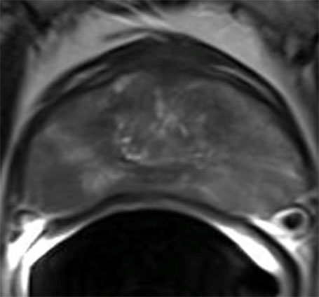
Axial T2
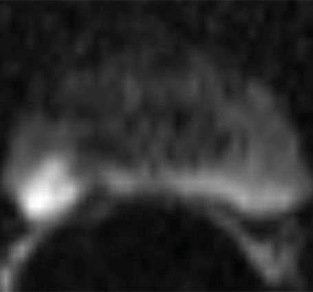
DWI
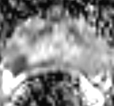
ADC Map
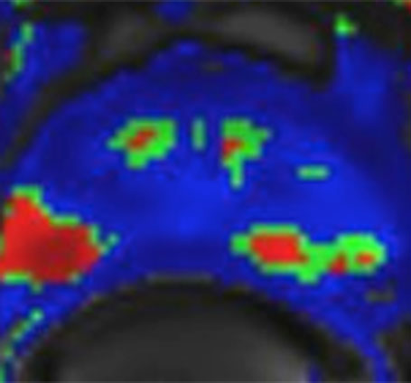
DCE
Case courtesy of Rajan T. Gupta, MD; Duke University Medical Center
3.0T Case Study 3 – 61 year old male, elevated PSA OF 8.12 ng/mLL
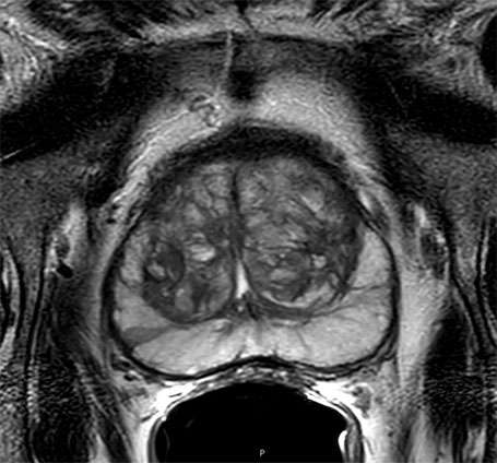
Axial T2
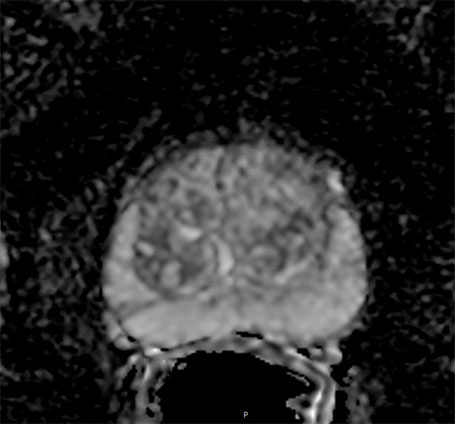
DWI
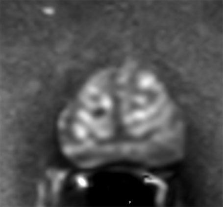
ADC Map
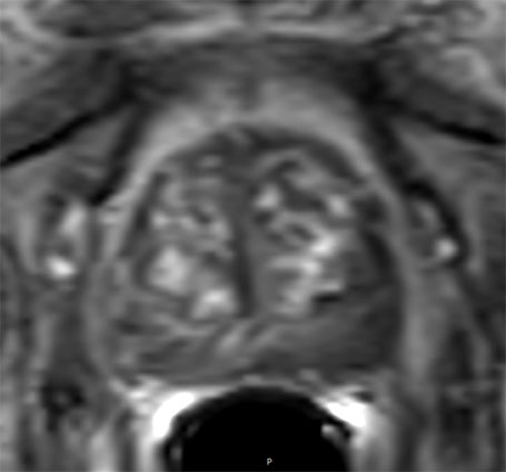
DcE
Case courtesy of Dr. Baris Turkbey and Dr. Peter Choyke, Molecular Imaging Branch, National Cancer Institute, NIH
3.0T Case Study 4 – 67 year old male, elevated PSA of 15.88 ng/mL
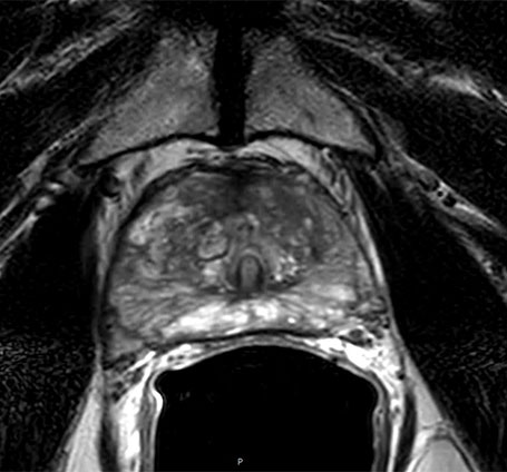
Axial T2
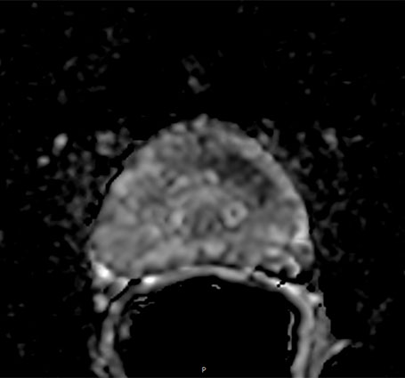
DWI
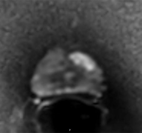
ADC Map
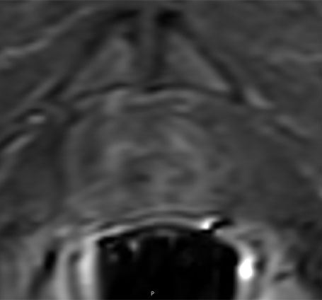
DcE
Case courtesy of Dr. Baris Turkbey and Dr. Peter Choyke, Molecular Imaging Branch, National Cancer Institute, NIH
3.0T Case Study 5
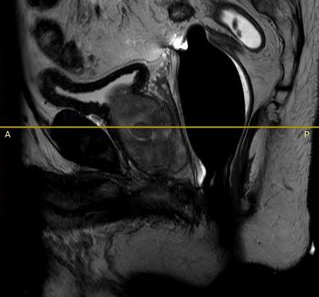
T2 Sagittal
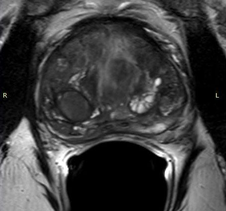
Axial T2
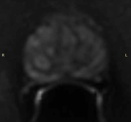
DWI
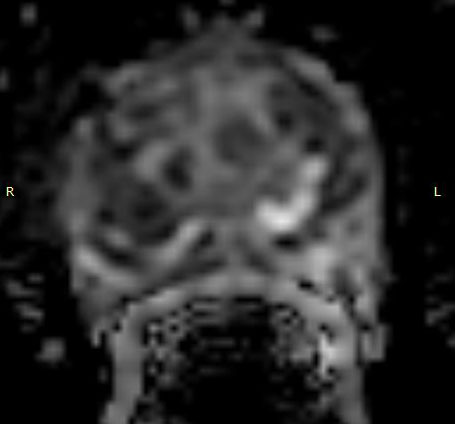
ADC Map
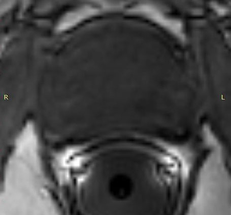
DCE
Case courtesy of Advanced Diagnostic Radiology – Cumberland, MD
3.0T Case Study 6
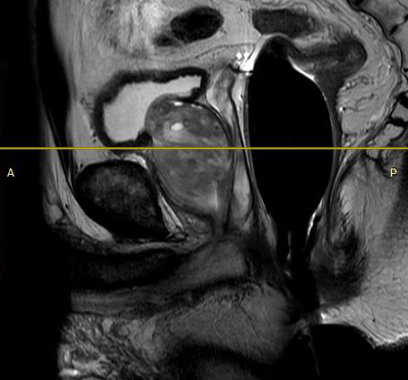
T2 Sagittal
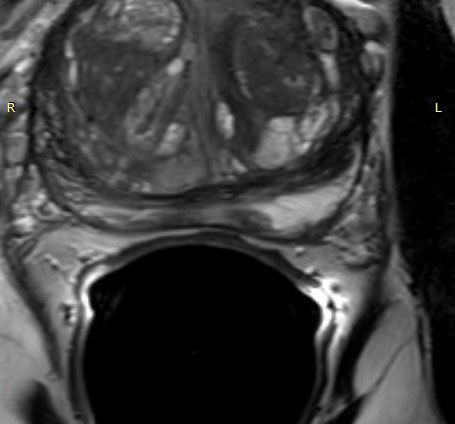
Axial T2
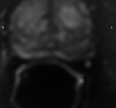
DWI
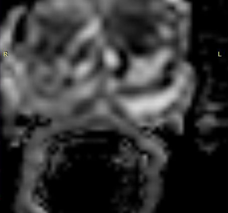
ADC Map
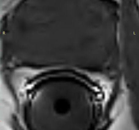
DCE
Case courtesy of Advanced Diagnostic Radiology – Cumberland, MD
3.0T Case Study 7
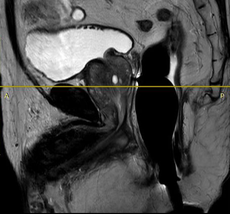
T2 Sagittal
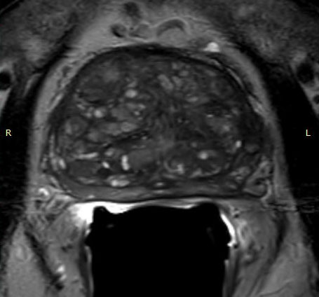
Axial T2
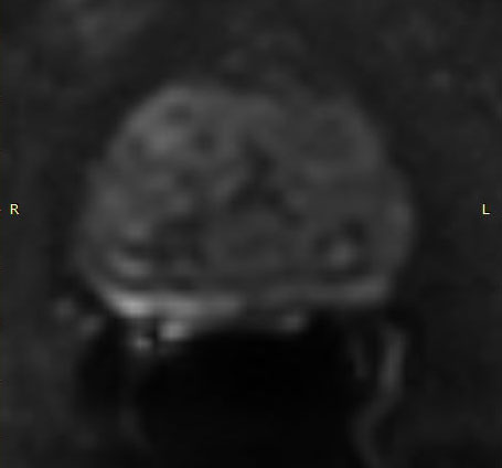
DWI
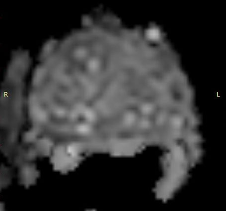
ADC Map
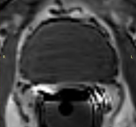
DCE
Case courtesy of Advanced Diagnostic Radiology – Cumberland, MD
3.0T Case Study 8
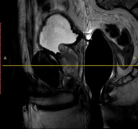
T2 Sagittal
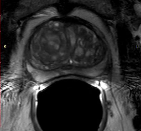
Axial T2
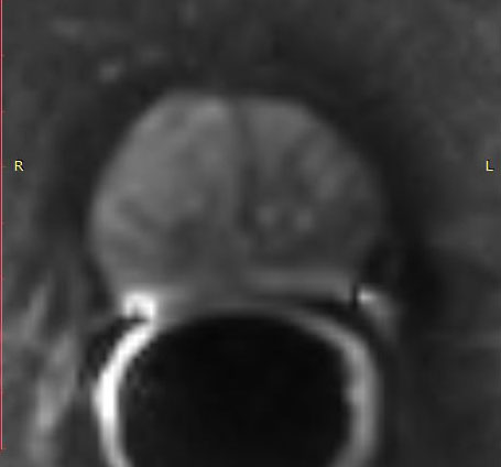
DWI
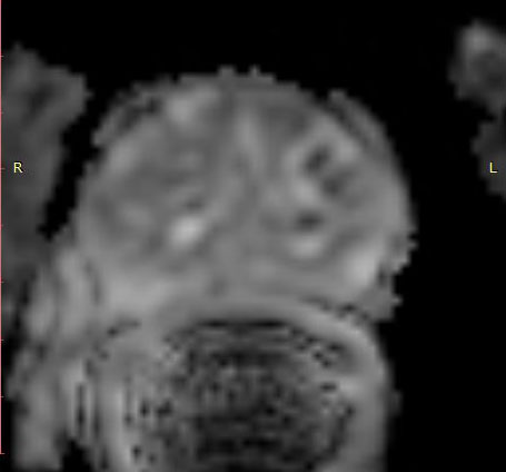
ADC Map
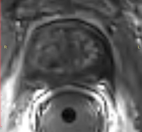
DCE
Case courtesy of Advanced Diagnostic Radiology – Cumberland, MD


